- Polymerase chain reaction, or PCR, is a technique to make many copies of a specific DNA region in vitro (in a test tube rather than an organism).
- PCR relies on a thermostable DNA polymerase, Taq polymerase, and requires DNA primers designed specifically for the DNA region of interest.
- In PCR, the reaction is repeatedly cycled through a series of temperature changes, which allow many copies of the target region to be produced.
- PCR has many research and practical applications. It is routinely used in DNA cloning, medical diagnostics, and forensic analysis of DNA.
What is PCR?
Polymerase chain reaction (PCR) is a common laboratory technique used to make many copies (millions or billions!) of a particular region of DNA. This DNA region can be anything the experimenter is interested in. For example, it might be a gene whose function a researcher wants to understand, or a genetic marker used by forensic scientists to match crime scene DNA with suspects.
Typically, the goal of PCR is to make enough of the target DNA region that it can be analyzed or used in some other way. For instance, DNA amplified by PCR may be sent for sequencing, visualized by gel electrophoresis, or clonedinto a plasmid for further experiments.
PCR is used in many areas of biology and medicine, including molecular biology research, medical diagnostics, and even some branches of ecology.
Taq polymerase
Like DNA replication in an organism, PCR requires a DNA polymerase enzyme that makes new strands of DNA, using existing strands as templates. The DNA polymerase typically used in PCR is called Taq polymerase, after the heat-tolerant bacterium from which it was isolated (Thermus aquaticus).
T. aquaticus lives in hot springs and hydrothermal vents. Its DNA polymerase is very heat-stable and is most active around 70°C (a temperature at which a human or E. coli DNA polymerase would be nonfunctional). This heat-stability makes Taq polymerase ideal for PCR. As we'll see, high temperature is used repeatedly in PCR to denature the template DNA, or separate its strands.
PCR primers
Like other DNA polymerases, Taq polymerase can only make DNA if it's given a primer, a short sequence of nucleotides that provides a starting point for DNA synthesis. In a PCR reaction, the experimenter determines the region of DNA that will be copied, or amplified, by the primers she or he chooses.
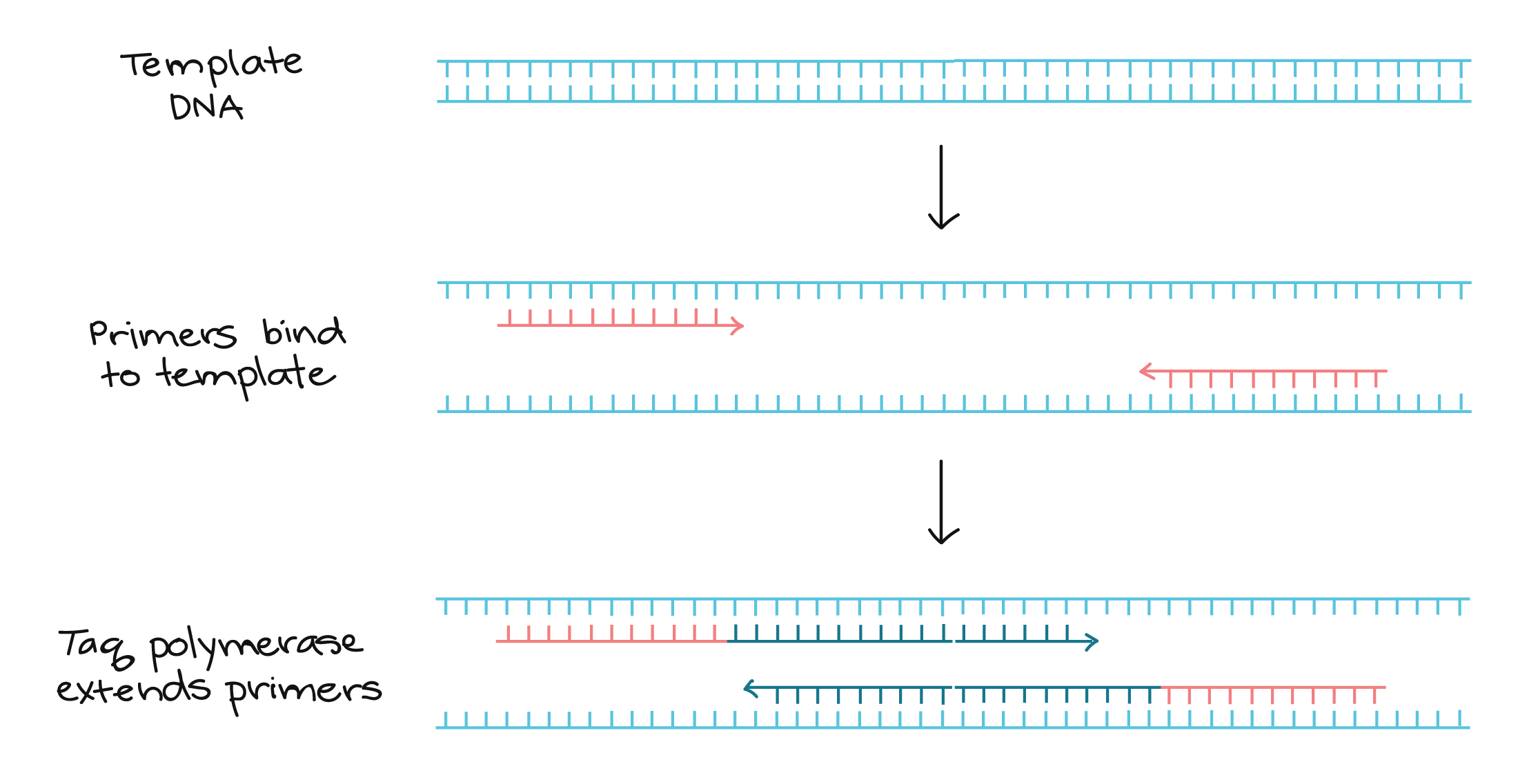
PCR primers are short pieces of single-stranded DNA, usually around 20nucleotides in length. Two primers are used in each PCR reaction, and they are designed so that they flank the target region (region that should be copied). That is, they are given sequences that will make them bind to opposite strands of the template DNA, just at the edges of the region to be copied. The primers bind to the template by complementary base pairing.

When the primers are bound to the template, they can be extended by the polymerase, and the region that lies between them will get copied.
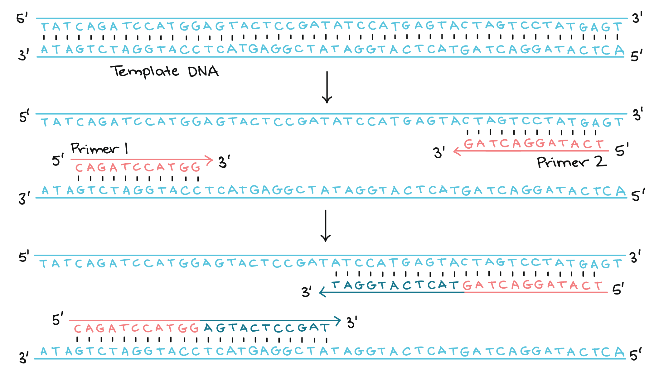
[More detailed diagram showing DNA and primer directionality]
The steps of PCR
The key ingredients of a PCR reaction are Taq polymerase, primers, template DNA, and nucleotides (DNA building blocks). The ingredients are assembled in a tube, along with cofactors needed by the enzyme, and are put through repeated cycles of heating and cooling that allow DNA to be synthesized.
The basic steps are:
- Denaturation (
96°C ): Heat the reaction strongly to separate, or denature, the DNA strands. This provides single-stranded template for the next step. - Annealing (55
- 65°C ): Cool the reaction so the primers can bind to their complementary sequences on the single-stranded template DNA. - Extension (
72°C ): Raise the reaction temperatures so Taq polymerase extends the primers, synthesizing new strands of DNA.

This cycle repeats 25 - 35 times in a typical PCR reaction, which generally takes 2 - 4 hours, depending on the length of the DNA region being copied. If the reaction is efficient (works well), the target region can go from just one or a few copies to billions.
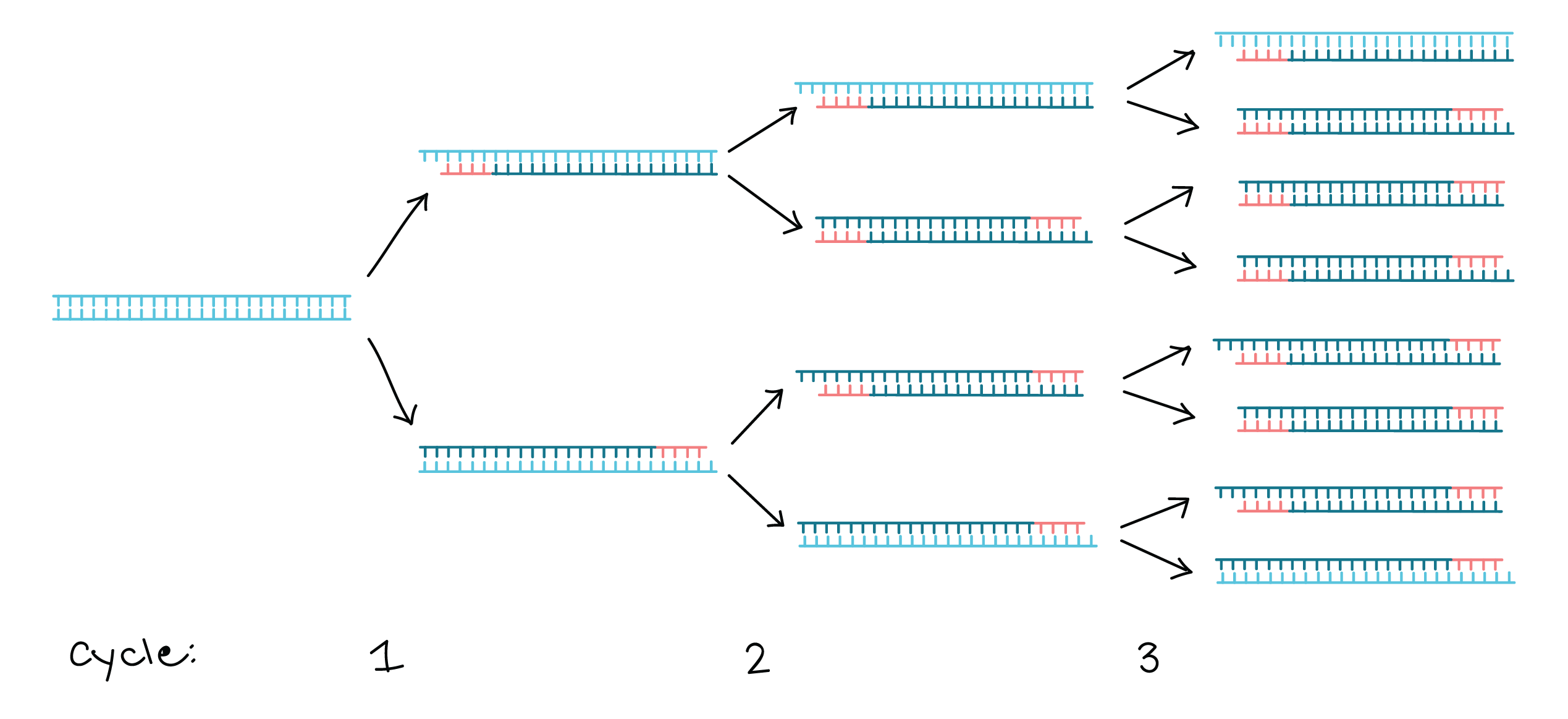
That’s because it’s not just the original DNA that’s used as a template each time. Instead, the new DNA that’s made in one round can serve as a template in the next round of DNA synthesis. There are many copies of the primers and many molecules of Taq polymerase floating around in the reaction, so the number of DNA molecules can roughly double in each round of cycling. This pattern of exponential growth is shown in the image below.
Using gel electrophoresis to visualize the results of PCR
The results of a PCR reaction are usually visualized (made visible) using gel electrophoresis. Gel electrophoresis is a technique in which fragments of DNA are pulled through a gel matrix by an electric current, and it separates DNA fragments according to size. A standard, or DNA ladder, is typically included so that the size of the fragments in the PCR sample can be determined.
DNA fragments of the same length form a "band" on the gel, which can be seen by eye if the gel is stained with a DNA-binding dye. For example, a PCR reaction producing a 400 base pair (bp) fragment would look like this on a gel:

A DNA band contains many, many copies of the target DNA region, not just one or a few copies. Because DNA is microscopic, lots of copies of it must be present before we can see it by eye. This is a big part of why PCR is an important tool: it produces enough copies of a DNA sequence that we can see or manipulate that region of DNA.
Applications of PCR
Using PCR, a DNA sequence can be amplified millions or billions of times, producing enough DNA copies to be analyzed using other techniques. For instance, the DNA may be visualized by gel electrophoresis, sent for sequencing, or digested with restriction enzymes and cloned into a plasmid.
PCR is used in many research labs, and it also has practical applications in forensics, genetic testing, and diagnostics. For instance, PCR is used to amplify genes associated with genetic disorders from the DNA of patients (or from fetal DNA, in the case of prenatal testing). PCR can also be used to test for a bacterium or DNA virus in a patient's body: if the pathogen is present, it may be possible to amplify regions of its DNA from a blood or tissue sample.
Sample problem: PCR in forensics
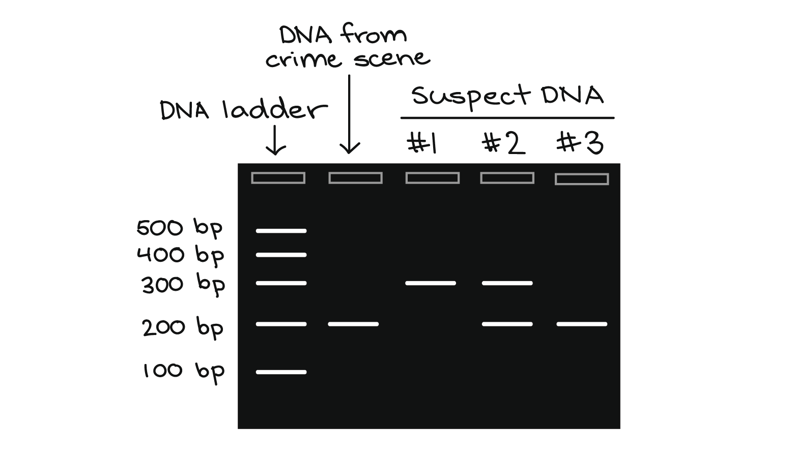
Suppose that you are working in a forensics lab. You have just received a DNA sample from a hair left at a crime scene, along with DNA samples from three possible suspects. Your job is to examine a particular genetic marker and see whether any of the three suspects matches the hair DNA for this marker.
The marker comes in two alleles, or versions. One contains a single repeat (brown region below), while the other contains two copies of the repeat. In a PCR reaction with primers that flank the repeat region, the first allele produces a 200 b, p DNA fragment, while the second produces a 300 b, p DNA fragment:

You perform PCR on the four DNA samples and visualize the results by gel electrophoresis, as shown below: