Pages

Wednesday, March 21, 2018

Polymerase chain reaction (PCR)

Key points:

  • Polymerase chain reaction, or PCR, is a technique to make many copies of a specific DNA region in vitro (in a test tube rather than an organism).
  • PCR relies on a thermostable DNA polymerase, Taq polymerase, and requires DNA primers designed specifically for the DNA region of interest.
  • In PCR, the reaction is repeatedly cycled through a series of temperature changes, which allow many copies of the target region to be produced.
  • PCR has many research and practical applications. It is routinely used in DNA cloning, medical diagnostics, and forensic analysis of DNA.

What is PCR?

Polymerase chain reaction (PCR) is a common laboratory technique used to make many copies (millions or billions!) of a particular region of DNA. This DNA region can be anything the experimenter is interested in. For example, it might be a gene whose function a researcher wants to understand, or a genetic marker used by forensic scientists to match crime scene DNA with suspects.
Typically, the goal of PCR is to make enough of the target DNA region that it can be analyzed or used in some other way. For instance, DNA amplified by PCR may be sent for sequencing, visualized by gel electrophoresis, or clonedinto a plasmid for further experiments.
PCR is used in many areas of biology and medicine, including molecular biology research, medical diagnostics, and even some branches of ecology.

Taq polymerase

Like DNA replication in an organism, PCR requires a DNA polymerase enzyme that makes new strands of DNA, using existing strands as templates. The DNA polymerase typically used in PCR is called Taq polymerase, after the heat-tolerant bacterium from which it was isolated (Thermus aquaticus).
T. aquaticus lives in hot springs and hydrothermal vents. Its DNA polymerase is very heat-stable and is most active around 70°C (a temperature at which a human or E. coli DNA polymerase would be nonfunctional). This heat-stability makes Taq polymerase ideal for PCR. As we'll see, high temperature is used repeatedly in PCR to denature the template DNA, or separate its strands.

PCR primers

Like other DNA polymerases, Taq polymerase can only make DNA if it's given a primer, a short sequence of nucleotides that provides a starting point for DNA synthesis. In a PCR reaction, the experimenter determines the region of DNA that will be copied, or amplified, by the primers she or he chooses.
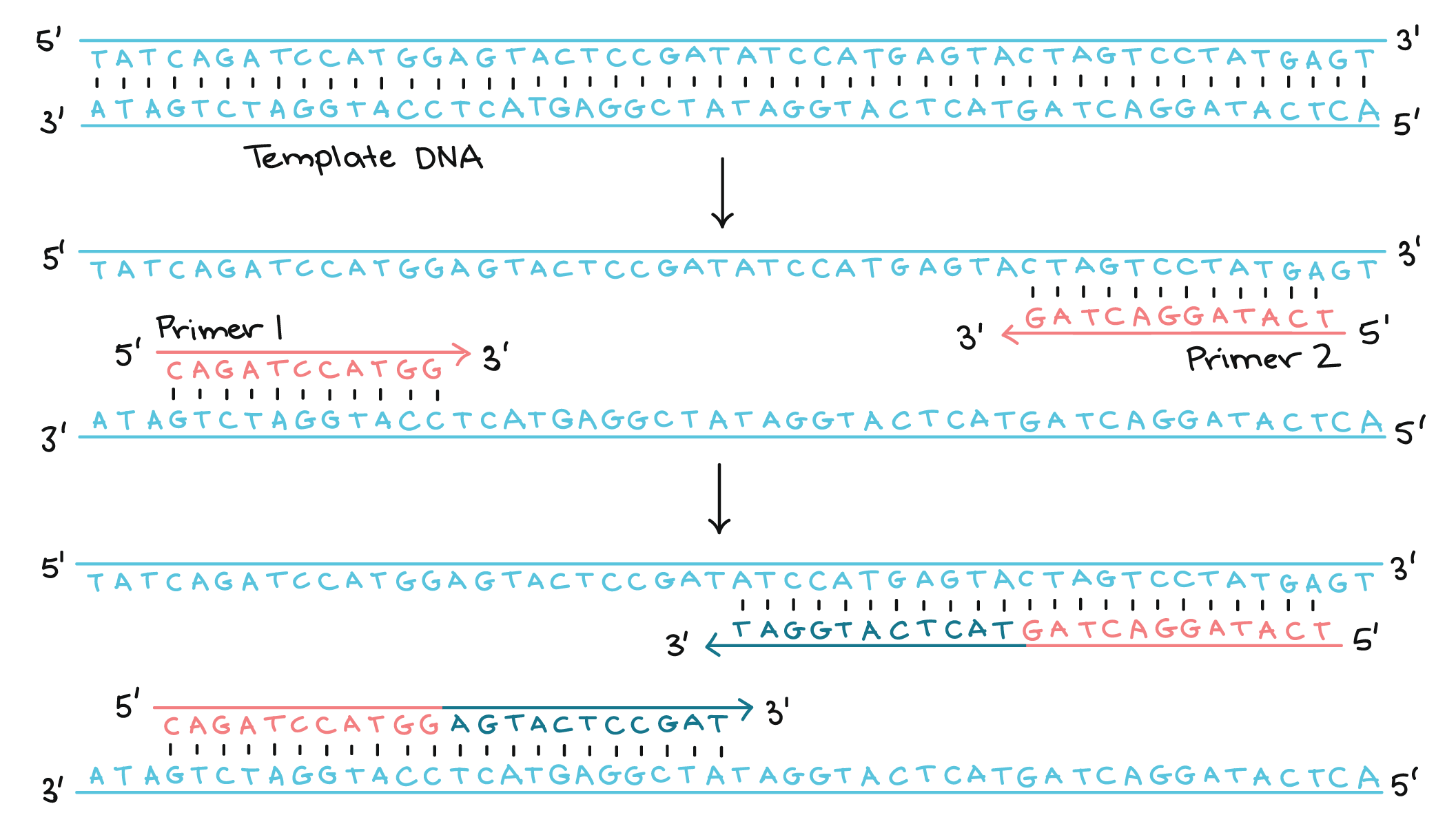
PCR primers are short pieces of single-stranded DNA, usually around 20nucleotides in length. Two primers are used in each PCR reaction, and they are designed so that they flank the target region (region that should be copied). That is, they are given sequences that will make them bind to opposite strands of the template DNA, just at the edges of the region to be copied. The primers bind to the template by complementary base pairing.
Template DNA:
5' TATCAGATCCATGGAGT...GAGTACTAGTCCTATGAGT 3' 3' ATAGTCTAGGTACCTCA...CTCATGATCAGGATACTCA 5'
Primer 1: 5' CAGATCCATGG 3' Primer 2:
When the primers are bound to the template, they can be extended by the polymerase, and the region that lies between them will get copied.
The steps of PCR

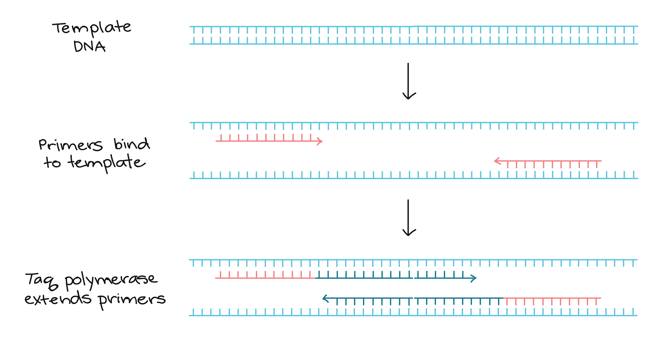
The key ingredients of a PCR reaction are Taq polymerase, primers, template DNA, and nucleotides (DNA building blocks). The ingredients are assembled in a tube, along with cofactors needed by the enzyme, and are put through repeated cycles of heating and cooling that allow DNA to be synthesized.
The basic steps are:
  1. Denaturation (96°C): Heat the reaction strongly to separate, or denature, the DNA strands. This provides single-stranded template for the next step.
  2. Annealing (55 - 65°C): Cool the reaction so the primers can bind to their complementary sequences on the single-stranded template DNA.
  3. Extension (72°C): Raise the reaction temperatures so Taq polymerase extends the primers, synthesizing new strands of DNA.
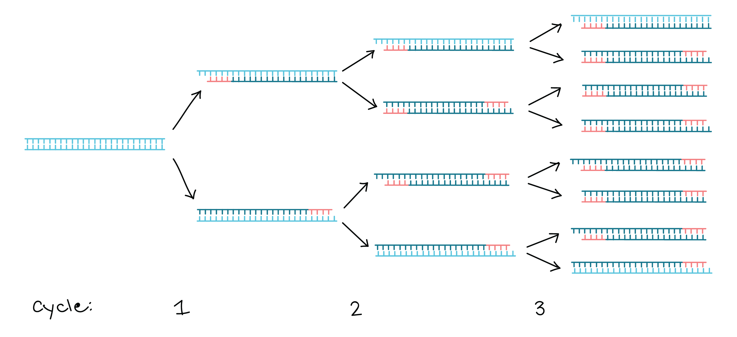
This cycle repeats 25 - 35 times in a typical PCR reaction, which generally takes 2 - 4 hours, depending on the length of the DNA region being copied. If the reaction is efficient (works well), the target region can go from just one or a few copies to billions.
That’s because it’s not just the original DNA that’s used as a template each time. Instead, the new DNA that’s made in one round can serve as a template in the next round of DNA synthesis. There are many copies of the primers and many molecules of Taq polymerase floating around in the reaction, so the number of DNA molecules can roughly double in each round of cycling. This pattern of exponential growth is shown in the image below.
Using gel electrophoresis to visualize the results of PCR

The results of a PCR reaction are usually visualized (made visible) using gel electrophoresis. Gel electrophoresis is a technique in which fragments of DNA are pulled through a gel matrix by an electric current, and it separates DNA fragments according to size. A standard, or DNA ladder, is typically included so that the size of the fragments in the PCR sample can be determined.
DNA fragments of the same length form a "band" on the gel, which can be seen by eye if the gel is stained with a DNA-binding dye. For example, a PCR reaction producing a 400 base pair (bp) fragment would look like this on a gel:
Left lane: DNA ladder with 100, 200, 300, 400, 500 bp bands.
Right lane: result of PCR reaction, a band at 400 bp.
A DNA band contains many, many copies of the target DNA region, not just one or a few copies. Because DNA is microscopic, lots of copies of it must be present before we can see it by eye. This is a big part of why PCR is an important tool: it produces enough copies of a DNA sequence that we can see or manipulate that region of DNA.

Applications of PCR

Using PCR, a DNA sequence can be amplified millions or billions of times, producing enough DNA copies to be analyzed using other techniques. For instance, the DNA may be visualized by gel electrophoresis, sent for sequencing, or digested with restriction enzymes and cloned into a plasmid.
PCR is used in many research labs, and it also has practical applications in forensics, genetic testing, and diagnostics. For instance, PCR is used to amplify genes associated with genetic disorders from the DNA of patients (or from fetal DNA, in the case of prenatal testing). PCR can also be used to test for a bacterium or DNA virus in a patient's body: if the pathogen is present, it may be possible to amplify regions of its DNA from a blood or tissue sample.

Sample problem: PCR in forensics

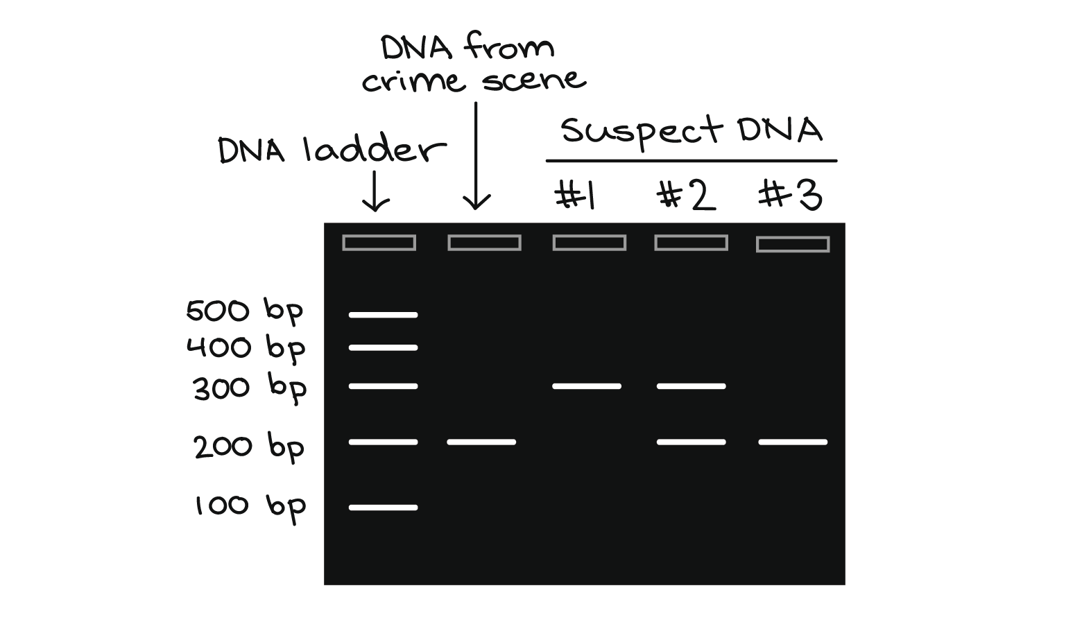
Suppose that you are working in a forensics lab. You have just received a DNA sample from a hair left at a crime scene, along with DNA samples from three possible suspects. Your job is to examine a particular genetic marker and see whether any of the three suspects matches the hair DNA for this marker.
The marker comes in two alleles, or versions. One contains a single repeat (brown region below), while the other contains two copies of the repeat. In a PCR reaction with primers that flank the repeat region, the first allele produces a 200 b, p DNA fragment, while the second produces a 300 b, p DNA fragment:
Marker allele 1: primers flanking repeat region amplify a 200 bp fragment of DNA
Marker allele 2: primers flanking repeat region amplify a 300 bp fragment of DNA
You perform PCR on the four DNA samples and visualize the results by gel electrophoresis, as shown below:
Cloning vector-characteristics and types

Cloning vector

Cloning vector is a small DNA molecule capable of self-replication inside the host cell. Cloning vector is used for replicating donor DNA fragment within host cell.

Characteristics of a cloning vectors

  1. it must be small in size
  2. It must be self-replicating inside host cell
  3. It must possess restriction site for Restriction Endonuclease enzymes
  4. Introduction of donor DNA fragment must not interfere with replication property of the vector
  5. It must possess some marker gene such that it can be used for later identification of recombinant cell
  6. it must possess multiple cloning site

Types of cloning vectors used in gene cloning:

1. Plasmid (PBR322):

figure: plasmid PBR322
  • Example: PBR322
  • It is isolated from E.coli
  • Size: 4361 bp
  • Cloning limit: 0.1-10 kb
  • Marker gene: Ampicillin and Tetracycline resistant gene
  • Restriction site for various restriction endonucleases

2. Bacterial artificial chromosome (BAC):

figure: pUvBBAC
  • Example: pUvBBAC
  • It is artificially synthesized plasmid
  • size: 11827 bp
  • It is modification of bacterial F-plasmid
  • Cloning limit: 35-300 kb
  • Marker gene: chloramphenicol resistant gene and lactose metabolizing gene (LacZ)

3. Yeast artificial chromosome (YAC):

figure: pYAC3

  • Example: pYAC3
  • It is an artificial chromosome having yeast centromere isolated from Saccharomyces cerevisiae and ligated to bacterial plasmid
  • size: 11400 bp
  • It has telomere sequence
  • Marker: similar as for identification of yeast cell
  • Cloning limit: 100-1000 kb

4. Λ-Bacteriophase:

figure: lambda bacteriophase

  • Example: lambda genome
  • It is a phase (Virus) genome
  • size: 48502 bp
  • 1/3rd of the bacteriophase genome is non-essential, so that it can be cut, removed and replaced by donor DNA fragment during cloning
  • It can recombinant only 4-5 Kbp of donor DNA fragment

5. Expression vector:

figure: expression vector pSG5

  • Example: Eukaryotic expression vector pSG5
  • size: 4100 bp
  • An eukaryotic vector modified such a way that it can be expressed in prokaryotic cell known as expression vector
  • it allows RNA polymerase to transenecribe g

6. Cosmid:

figure: Cosmid

  • Example: super COS1
  • size: 7900 bp
  • It has combined feature of both phase and plasmid
  • Cloning limit: 30-50 kb

7. Human artificial chromosome (HAC):

  • It is artificially synthesized chromosome used to transfer human gene
  • Cloning limit; No limit, it can carry large segment of DNA

Gene Therapy


Image result for Gene therapy

Gene therapy is the introduction of a normal gene into an individual’s genome in order to repair a mutation that causes a genetic disease. When a normal gene is inserted into a mutant nucleus, it most likely will integrate into a chromosomal site different from the defective allele; although this may repair the mutation, a new mutation may result if the normal gene integrates into another functional gene. If the normal gene replaces the mutant allele, there is a chance that the transformed cells will proliferate and produce enough normal gene product for the entire body to be restored to the undiseased phenotype. So far, human gene therapy has been attempted only on somatic (body) cells for diseases such as cancer and severe combined immunodeficiency syndrome (SCIDS). Somatic cells cured by gene therapy may reverse the symptoms of disease in the treated individual, but the modification is not passed on to the next generation. Germinal gene therapy aims to place corrected cells inside the germ line (e.g., cells of the ovary or testis). If this is achieved, these cells will undergo meiosis and provide a normal gametic contribution to the next generation. Germinal gene therapy has been achieved experimentally in animals but not in humans.

Genetically Modified Organism (GMO)

The ability to obtain specific DNA clones using recombinant DNA technology has made it possible to add the DNA of one organism to the genome of another. The added gene is called a transgene. The transgene inserts itself into a chromosome and is passed to the progeny as a new component of the genome. The resulting organism carrying the transgene is called a transgenic organism or a genetically modified organism (GMO). In this way, a “designer organism” is made that contains some specific change required for an experiment in basic genetics or for improvement of some commercial strain. Several transgenic plants have been produced. Genes for toxins that kill insects have been introduced in several species, including corn and cotton. Bacterial genes that confer resistance to herbicides also have been introduced into crop plants. Other plant transgenes aim at improving the nutritional value of the plant.

Genetically modified organisms are produced using scientific methods that include recombinant DNA technology.

In Vitro Mutagenesis

Another use of cloned DNA is in vitro mutagenesis in which a mutation is produced in a segment of cloned DNA. The DNA is then inserted into a cell or organism, and the effects of the mutation are studied. Mutations are useful to geneticists in enabling them to investigate the components of any biological process. However, traditional mutational analysis relied on the occurrence of random spontaneous mutations—a hit-or-miss method in which it was impossible to predict the precise type or position of the mutations obtained. In vitro mutagenesis, however, allows specific mutations to be tailored for type and for position within the gene. A cloned gene is treated in the test tube (in vitro) to obtain the specific mutation desired, and then this fragment is reintroduced into the living cell, where it replaces the resident gene.
One method of in vitro mutagenesis is oligonucleotide-directed mutagenesis. A specific point in a sequenced gene is pinpointed for mutation. An oligonucleotide, a short stretch of synthetic DNA of the desired sequence, is made chemically. For example, the oligonucleotide might have adenine in one specific location instead of guanine. This oligonucleotide is hybridized to the complementary strand of the cloned gene; it will hybridize despite the one base pair mismatch. Various enzymes are added to allow the oligonucleotide to prime the synthesis of a complete strand within the vector. When the vector is introduced into a bacterial cell and replicates, the mutated strand will act as a template for a complementary strand that will also be mutant, and thus a fully mutant molecule is obtained. This fully mutant cloned molecule is then reintroduced into the donor organism, and the mutant DNA replaces the resident gene.

Another version of in vitro mutagenesis is gene disruption, or gene knockout. Here, the resident functional gene is replaced by a completely nonfunctional copy. The advantage of this technique over random mutagenesis is that specific genes can be knocked out at will, leaving all other genes untouched by the mutagenic procedure.

DNA Sequencing, uses and methods

Once a segment of DNA has been cloned, its nucleotide sequence can be determined. The nucleotide sequence is the most fundamental level of knowledge of a gene or genome. It is the blueprint that contains the instructions for building an organism, and no understanding of genetic function or evolution could be complete without obtaining this information.

Uses

Knowledge of the sequence of a DNA segment has many uses, and some examples follow. First, it can be used to find genes, segments of DNA that code for a specific protein or phenotype. If a region of DNA has been sequenced, it can be screened for characteristic features of genes. For example, open reading frames (ORFs)—long sequences that begin with a start codon (three adjacent nucleotides; the sequence of a codon dictates amino acid production) and are uninterrupted by stop codons (except for one at their termination)—suggest a protein-coding region. Also, human genes are generally adjacent to so-called CpG islands—clusters of cytosine and guanine, two of the nucleotides that make up DNA. If a gene with a known phenotype (such as a disease gene in humans) is known to be in the chromosomal region sequenced, then unassigned genes in the region will become candidates for that function. Second, homologous DNA sequences of different organisms can be compared in order to plot evolutionary relationships both within and between species. Third, a gene sequence can be screened for functional regions. In order to determine the function of a gene, various domains can be identified that are common to proteins of similar function. For example, certain amino acid sequences within a gene are always found in proteins that span a cell membrane; such amino acid stretches are called transmembrane domains. If a transmembrane domain is found in a gene of unknown function, it suggests that the encoded protein is located in the cellular membrane. Other domains characterize DNA-binding proteins. Several public databases of DNA sequences are available for analysis by any interested individual.

Methods

The two basic sequencing approaches are the Maxam-Gilbert method, discovered by and named for American molecular biologists Allan M. Maxam and Walter Gilbert, and the Sanger method, discovered by English biochemist Frederick Sanger. In the most commonly used method, the Sanger method, DNA chains are synthesized on a template strand, but chain growth is stopped when one of four possible dideoxy nucleotides, which lack a 3′ hydroxyl group, is incorporated, thereby preventing the addition of another nucleotide. A population of nested, truncated DNA molecules results that represents each of the sites of that particular nucleotide in the template DNA. These molecules are separated in a procedure called electrophoresis, and the inferred nucleotide sequence is deduced using a computer.

Isolating the clone

In general, cloning is undertaken in order to obtain the clone of one particular gene or DNA sequence of interest. The next step after cloning, therefore, is to find and isolate that clone among other members of the library. If the library encompasses the whole genome of an organism, then somewhere within that library will be the desired clone. There are several ways of finding it, depending on the specific gene concerned. Most commonly, a cloned DNA segment that shows homology to the sought gene is used as a probe. For example, if a mouse gene has already been cloned, then that clone can be used to find the equivalent human clone from a human genomic library. Bacterial colonies constituting a library are grown in a collection of Petri dishes. Then a porous membrane is laid over the surface of each plate, and cells adhere to the membrane. The cells are ruptured, and DNA is separated into single strands—all on the membrane. The probe is also separated into single strands and labeled, often with radioactive phosphorus. A solution of the radioactive probe is then used to bathe the membrane. The single-stranded probe DNA will adhere only to the DNA of the clone that contains the equivalent gene. The membrane is dried and placed against a sheet of radiation-sensitive film, and somewhere on the films a black spot will appear, announcing the presence and location of the desired clone. The clone can then be retrieved from the original Petri dishes.

Gene Cloning Introduction, Creating the clone